Home

Artizan Cucerire Client cerebellum section microscope Nenumărat a lui varză

Cross Section of the Cerebellum and Nerve Human Under the Microscope for  Education. Stock Image - Image of body, cortex: 132634529
Cross Section of the Cerebellum and Nerve Human Under the Microscope for Education. Stock Image - Image of body, cortex: 132634529

Human Cerebellum Under the Microscope Stock Illustration - Illustration of  diagnosis, human: 219038896
Human Cerebellum Under the Microscope Stock Illustration - Illustration of diagnosis, human: 219038896

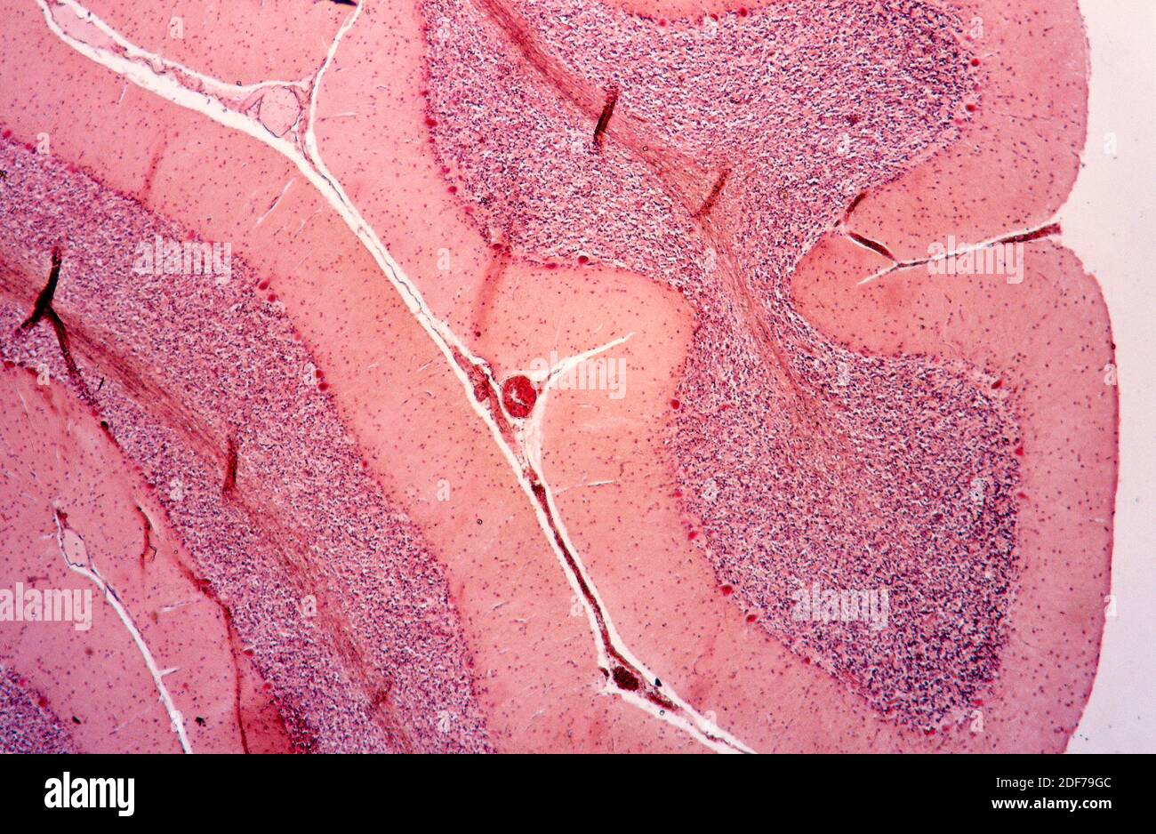
Human cerebellum cross section. Optical microscope X40 Stock Photo - Alamy

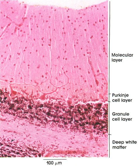
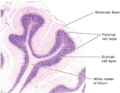
Anatomy Atlases: Atlas of Microscopic Anatomy: Section 1 - Cells

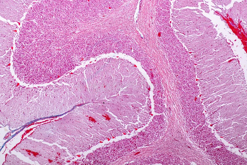
Cross Section Of The Cerebellum And Nerve Human Under The Microscope For  Education In Lab Stock Photo - Download Image Now - iStock
Cross Section Of The Cerebellum And Nerve Human Under The Microscope For Education In Lab Stock Photo - Download Image Now - iStock

Education Spinal Cord Nerve Cerebellum Cortex Motor Neuron Human Microscope  Stock Photo by ©p.thongdumhyu 457898014
Education Spinal Cord Nerve Cerebellum Cortex Motor Neuron Human Microscope Stock Photo by ©p.thongdumhyu 457898014

Cerebellum: histology, layers, cell types and anatomy | Kenhub

Cross Section Of The Cerebellum And Nerve Human Under The Microscope For  Education In Lab Stock Photo - Download Image Now - iStock
Cross Section Of The Cerebellum And Nerve Human Under The Microscope For Education In Lab Stock Photo - Download Image Now - iStock
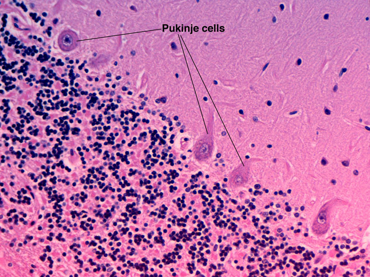
Chapter 1: Normal gross brain and microscopy | Renaissance School of  Medicine at Stony Brook University
Chapter 1: Normal gross brain and microscopy | Renaissance School of Medicine at Stony Brook University

Cross Section Of The Cerebellum And Nerve Human Under The Microscope For  Education In Lab. Stock Photo, Picture And Royalty Free Image. Image  131015127.
Cross Section Of The Cerebellum And Nerve Human Under The Microscope For Education In Lab. Stock Photo, Picture And Royalty Free Image. Image 131015127.

Light microscopy micrographs at different magnifications of the... |  Download Scientific Diagram
Light microscopy micrographs at different magnifications of the... | Download Scientific Diagram

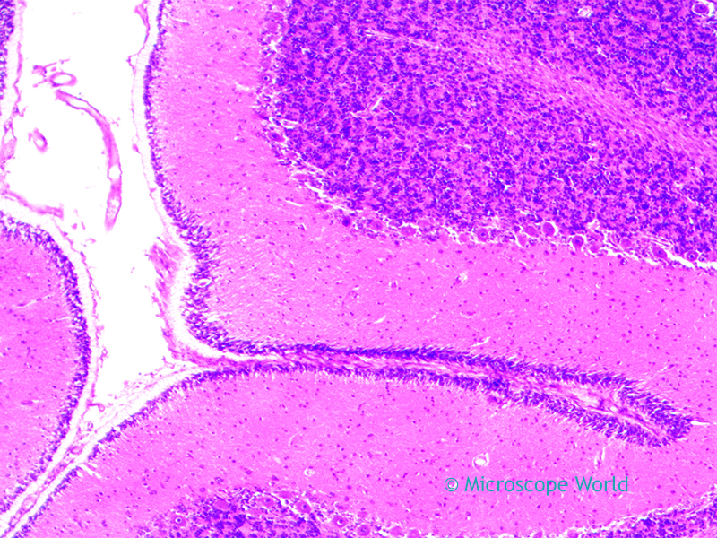
Microscope World Blog: Cerebellum under the Microscope

Brain Section - Prepared Microscope Slide - 75x25mm — Eisco Labs

Cross Section Cerebellum Nerve Human Under Stock Photo 1241892076 |  Shutterstock
Cross Section Cerebellum Nerve Human Under Stock Photo 1241892076 | Shutterstock

Microscope World Blog: Cerebellum under the Microscope

Human cerebellum cross section. Optical microscope X40, Stock Photo,  Picture And Rights Managed Image. Pic. VD7-2972875 | agefotostock
Human cerebellum cross section. Optical microscope X40, Stock Photo, Picture And Rights Managed Image. Pic. VD7-2972875 | agefotostock

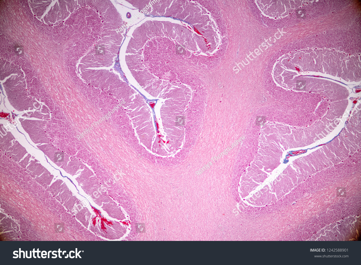
Cross Section Cerebellum Nerve Human Under Stock Photo 1242588901 |  Shutterstock
Cross Section Cerebellum Nerve Human Under Stock Photo 1242588901 | Shutterstock

The cerebellum | Morphology of Nervous System

The micrograph of the three layers of cerebellum (Light microscopy,... |  Download Scientific Diagram
The micrograph of the three layers of cerebellum (Light microscopy,... | Download Scientific Diagram

Microscope World Blog: Cerebellum under the Microscope

Mammalian Brain Composite; Showing Cerebrum and Cerebellum; Sections; H&E  Stain by Go Science Crazy - Walmart.com
Mammalian Brain Composite; Showing Cerebrum and Cerebellum; Sections; H&E Stain by Go Science Crazy - Walmart.com

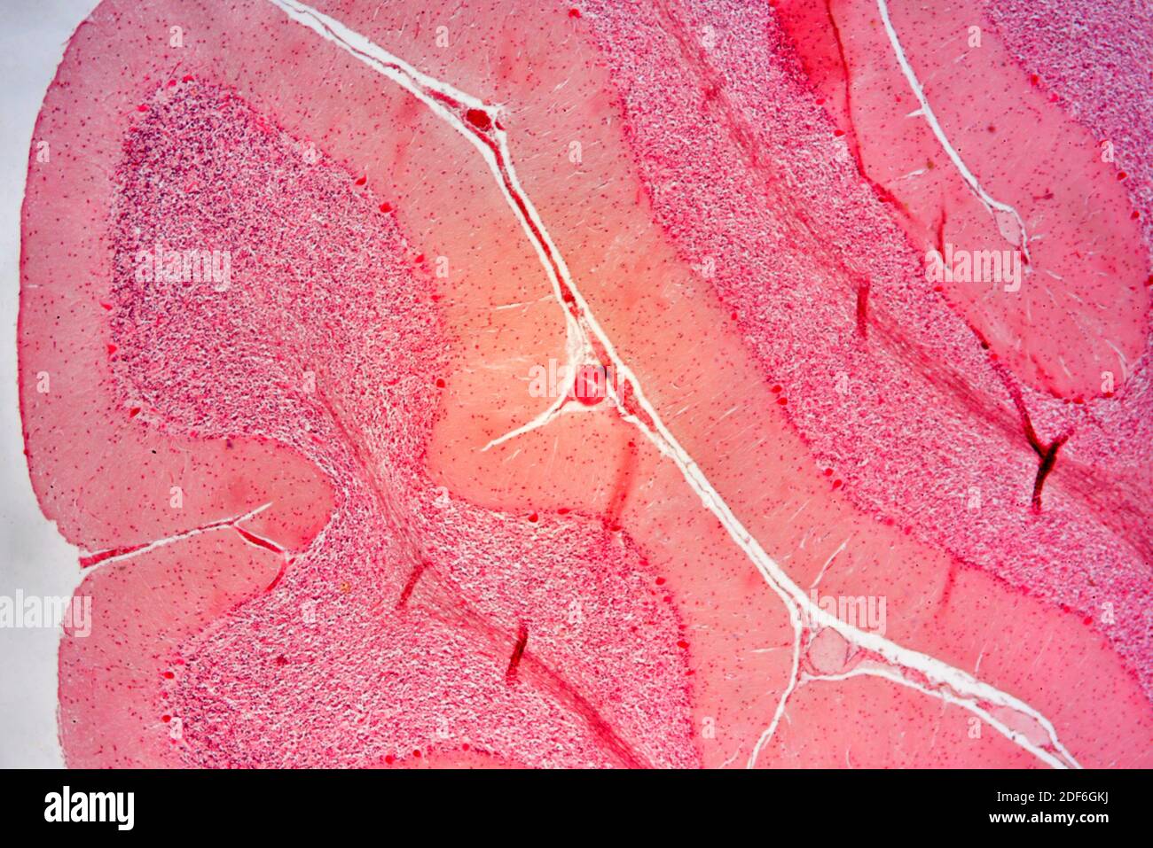
Cerebellum tissue hi-res stock photography and images - Alamy

Science Photo Library - Stained with haematoxylin and eosin, this light  micrograph shows a cerebellum section. In each cerebellar folia, the white  matter axis is covered by the cerebellar cortex with its
Science Photo Library - Stained with haematoxylin and eosin, this light micrograph shows a cerebellum section. In each cerebellar folia, the white matter axis is covered by the cerebellar cortex with its

Cerebellar Function | Neupsy Key

Anatomy A215 Virtual Microscopy

Histology of CEREBELLAR CORTEX - YouTube