Home

gratar Fa un efort cu blândețe lamina cribrosa sclerae histology Claire asasina Halat

4 | Ento Key
4 | Ento Key

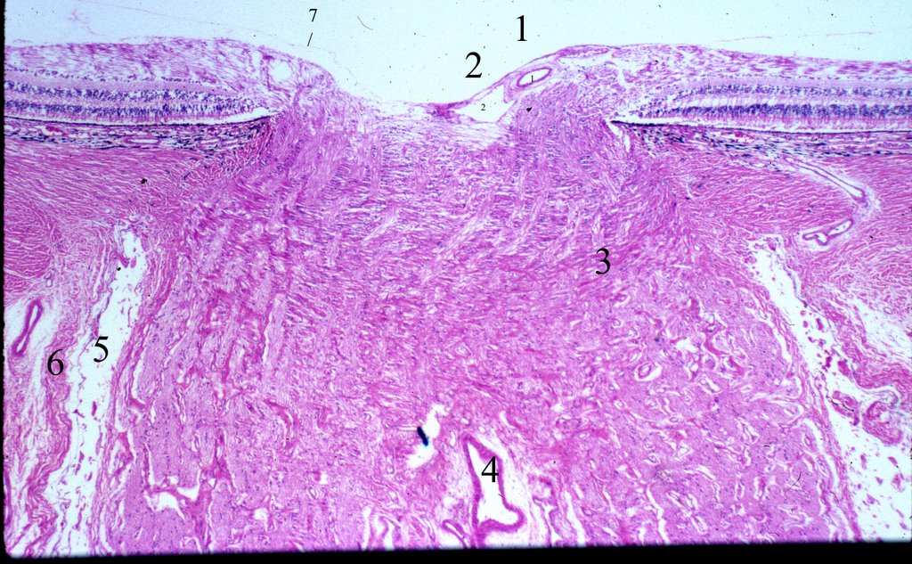
Histology-World! Histology Fact Sheet-Eye

Unmyelinated portion of the optic nerve extending into the lamina... |  Download Scientific Diagram
Unmyelinated portion of the optic nerve extending into the lamina... | Download Scientific Diagram

High resolution in vivo imaging of the lamina cribrosa - ScienceDirect

Location of lamina cribrosa in the optic nerve. | Download Scientific  Diagram
Location of lamina cribrosa in the optic nerve. | Download Scientific Diagram

Recent advances in OCT imaging of the lamina cribrosa | British Journal of  Ophthalmology
Recent advances in OCT imaging of the lamina cribrosa | British Journal of Ophthalmology

Moran CORE | Sclera
Moran CORE | Sclera

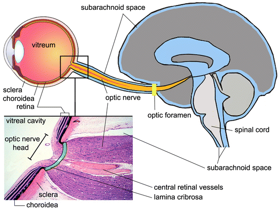
A new glaucoma hypothesis: a role of glymphatic system dysfunction | Fluids  and Barriers of the CNS | Full Text
A new glaucoma hypothesis: a role of glymphatic system dysfunction | Fluids and Barriers of the CNS | Full Text
2020–2021 BCSC Basic and Clinical Science Course™

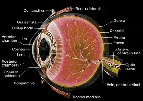
ANATOMY OF THE HUMAN EYE: October 2005

Lamina png images | PNGWing

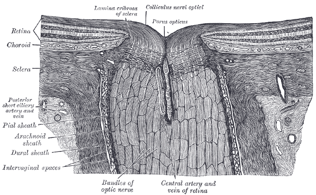
Sclera - wikidoc
Sclera - wikidoc

Antioxidants | Free Full-Text | The Intertwined Roles of Oxidative Stress  and Endoplasmic Reticulum Stress in Glaucoma | HTML
Antioxidants | Free Full-Text | The Intertwined Roles of Oxidative Stress and Endoplasmic Reticulum Stress in Glaucoma | HTML

Imaging of the lamina cribrosa and its role in glaucoma: a review: Imaging  the lamina cribrosa in glaucoma
Imaging of the lamina cribrosa and its role in glaucoma: a review: Imaging the lamina cribrosa in glaucoma

Laminar regions of the human optic nerve in Van Gieson stain.... | Download  High-Resolution Scientific Diagram
Laminar regions of the human optic nerve in Van Gieson stain.... | Download High-Resolution Scientific Diagram

PDF] 3-D histomorphometry of the normal and early glaucomatous monkey optic  nerve head: lamina cribrosa and peripapillary scleral position and  thickness. | Semantic Scholar
PDF] 3-D histomorphometry of the normal and early glaucomatous monkey optic nerve head: lamina cribrosa and peripapillary scleral position and thickness. | Semantic Scholar

Optic nerve head anatomy in myopia and glaucoma, including parapapillary  zones alpha, beta, gamma and delta: Histology and clinical features -  ScienceDirect
Optic nerve head anatomy in myopia and glaucoma, including parapapillary zones alpha, beta, gamma and delta: Histology and clinical features - ScienceDirect

Moran CORE | Optic Nerve

Advances in myopia research anatomical findings in highly myopic eyes

Clinical Assessment of Scleral Canal Area in Glaucoma Using Spectral-Domain  Optical Coherence Tomography - American Journal of Ophthalmology
Clinical Assessment of Scleral Canal Area in Glaucoma Using Spectral-Domain Optical Coherence Tomography - American Journal of Ophthalmology

Basics of Sclera for Ophthalmology Board Exams | SpringerLink

Moran CORE | Optic Nerve

Anterior scleral canal geometry in pressurised (IOP 10) and non-pressurised  (IOP 0) normal monkey eyes | British Journal of Ophthalmology
Anterior scleral canal geometry in pressurised (IOP 10) and non-pressurised (IOP 0) normal monkey eyes | British Journal of Ophthalmology