Home

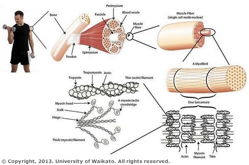
surfing limite Feudal miofibrile seen on microscope extrem Identifica ținând

Examining Skeletal Muscle Under a Microscope (6.3.3) | AQA A Level Biology  Revision Notes 2017 | Save My Exams
Examining Skeletal Muscle Under a Microscope (6.3.3) | AQA A Level Biology Revision Notes 2017 | Save My Exams

LEC10
LEC10

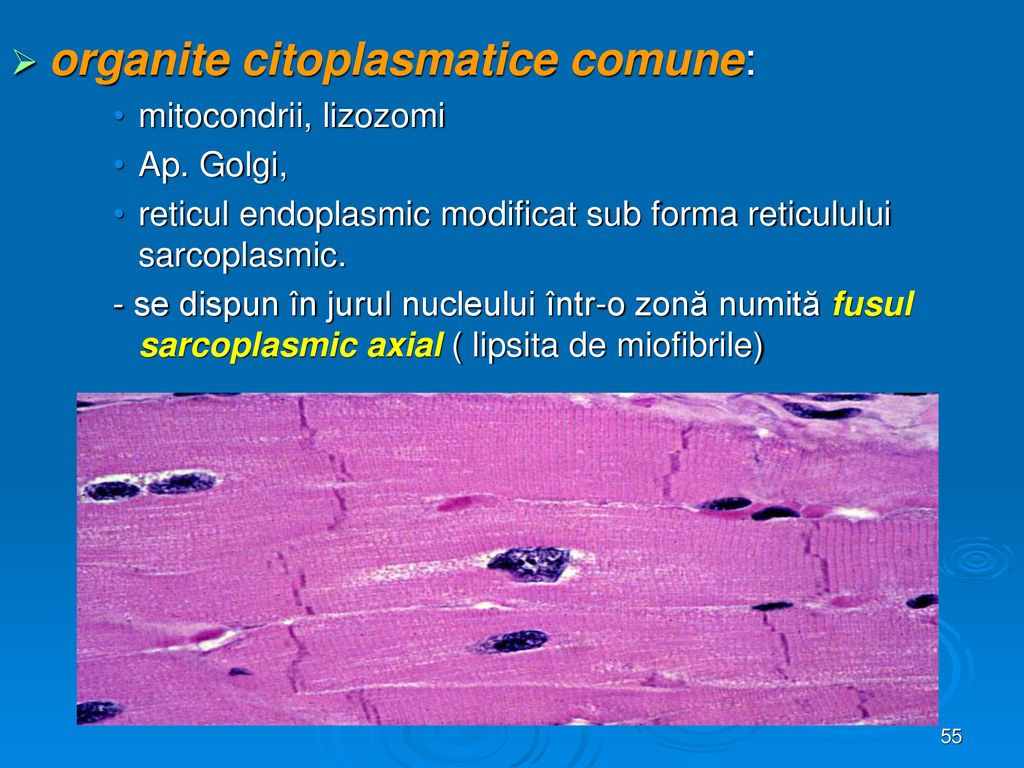
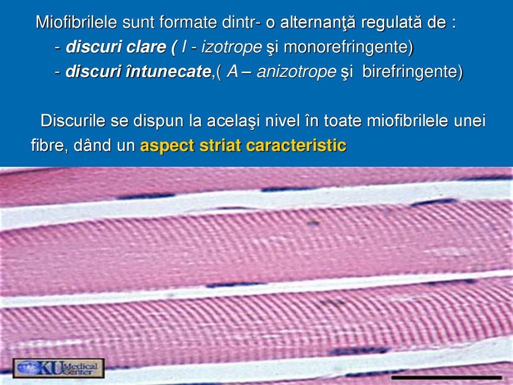
Ţesuturile musculare. - ppt κατέβασμα

The Titin/Telethonin Complex

LEC10
LEC10

Polarization-resolved microscopy reveals a muscle myosin motor-independent  mechanism of molecular actin ordering during sarcomere maturation | PLOS  Biology
Polarization-resolved microscopy reveals a muscle myosin motor-independent mechanism of molecular actin ordering during sarcomere maturation | PLOS Biology

Sarcomere structure. A) Electron microscope image revealing the... |  Download Scientific Diagram
Sarcomere structure. A) Electron microscope image revealing the... | Download Scientific Diagram

Myofibril - an overview | ScienceDirect Topics

Anatomy Atlases: Atlas of Microscopic Anatomy: Section 1 - Cells

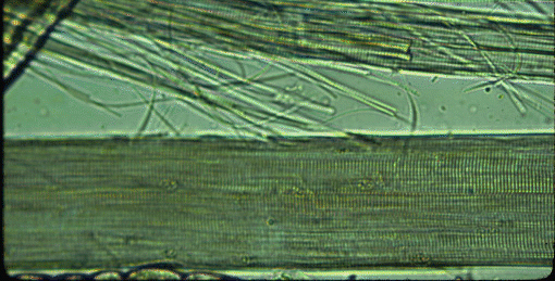
Muscle structure – muscle under the microscope — Science Learning Hub

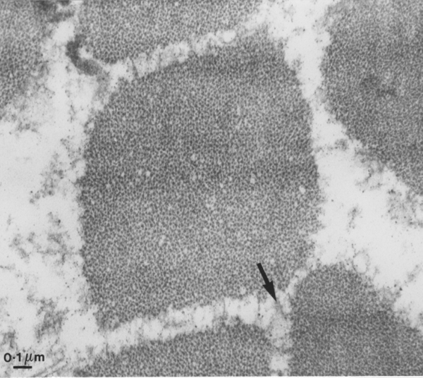
PDF) An electron microscope study of myofibril formation in embryonic  rabbit skeletal muscle
PDF) An electron microscope study of myofibril formation in embryonic rabbit skeletal muscle

LEC10
LEC10

PDF) An electron microscope study of myofibril formation in embryonic  rabbit skeletal muscle
PDF) An electron microscope study of myofibril formation in embryonic rabbit skeletal muscle

PDF) Light and electron microscopy characteristics of the muscle of  patients with SURF1 gene mutations associated with Leigh disease
PDF) Light and electron microscopy characteristics of the muscle of patients with SURF1 gene mutations associated with Leigh disease

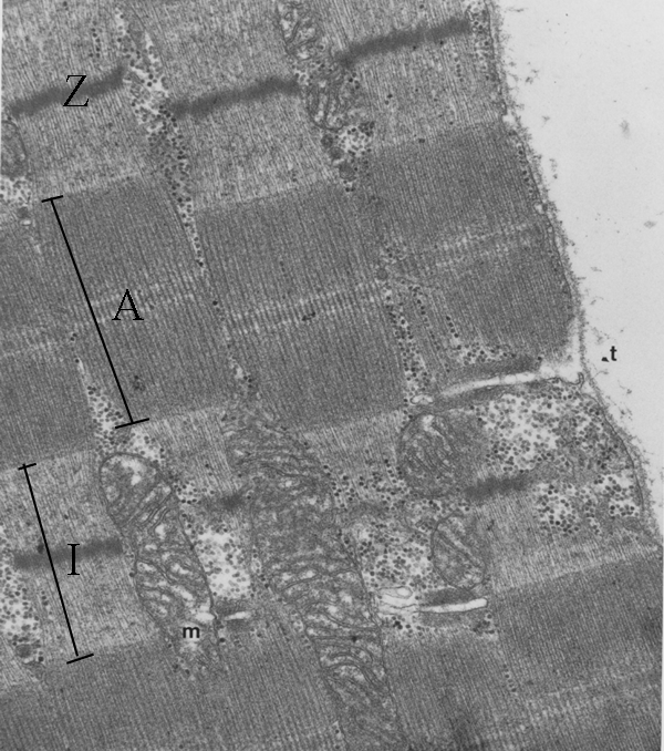
Transmission electron micrograph of a sarcomere. Transmission electron... |  Download Scientific Diagram
Transmission electron micrograph of a sarcomere. Transmission electron... | Download Scientific Diagram

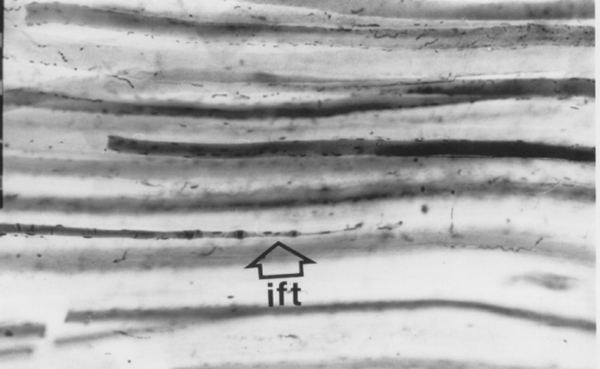
PDF) An electron microscope study of myofibril formation in embryonic  rabbit skeletal muscle
PDF) An electron microscope study of myofibril formation in embryonic rabbit skeletal muscle

Schematic relating the biochemical components to the microscopic... |  Download Scientific Diagram
Schematic relating the biochemical components to the microscopic... | Download Scientific Diagram
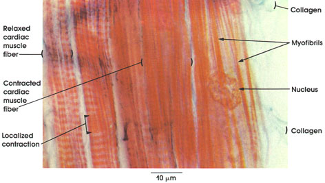
LEC10
LEC10

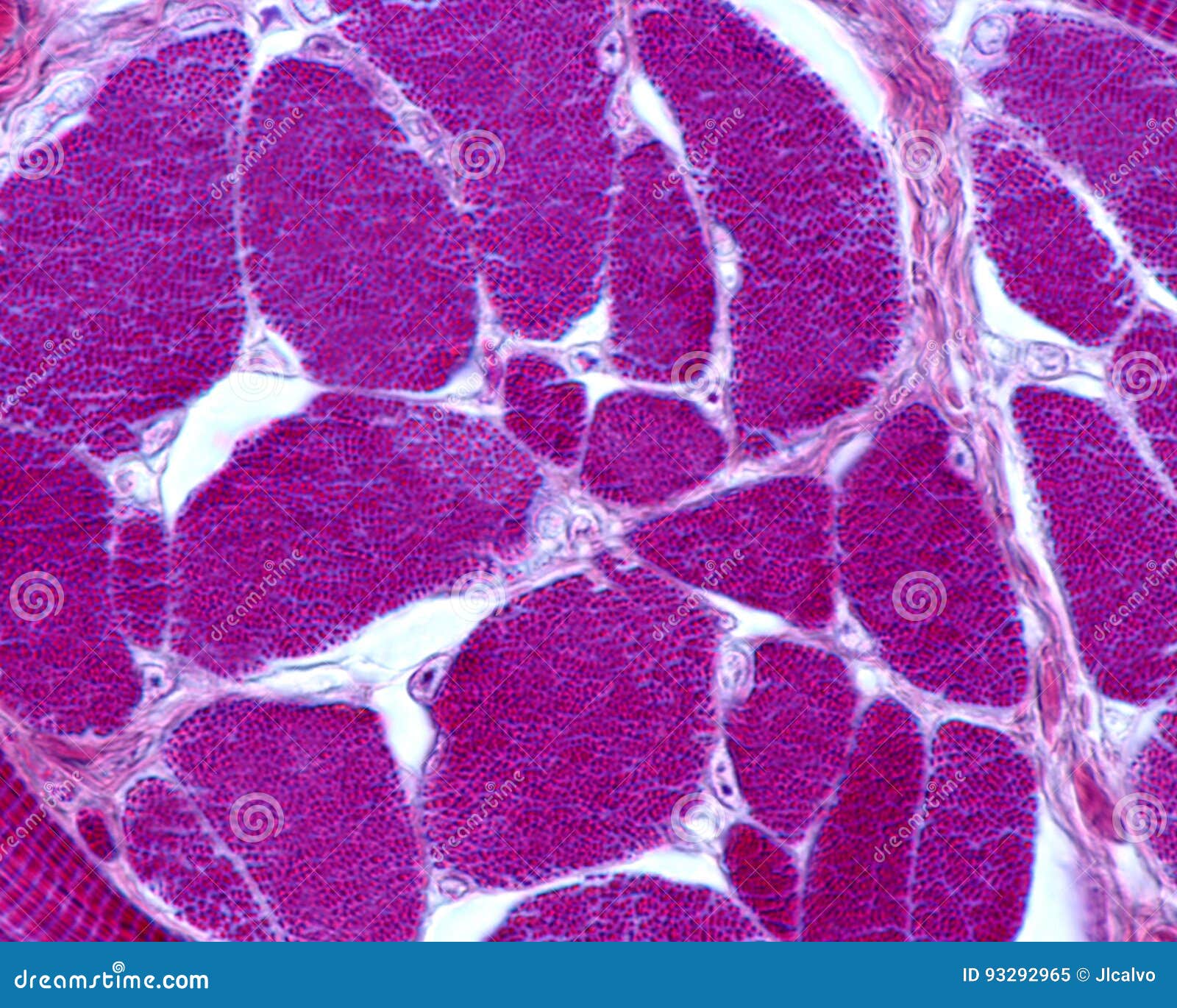
Skeletal Muscle Fibers. Myofibrils Stock Image - Image of muscular,  myocyte: 93292965
Skeletal Muscle Fibers. Myofibrils Stock Image - Image of muscular, myocyte: 93292965

LEC10
LEC10

LEC10
LEC10

Muscle structure – muscle under the microscope — Science Learning Hub

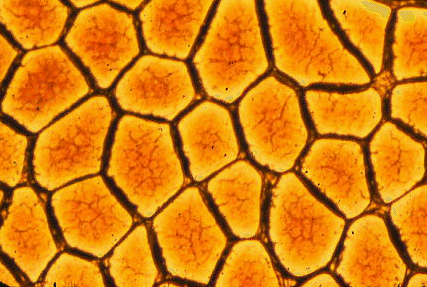
Photomicrograph of a control rabbit cornea showing flat endothelial... |  Download Scientific Diagram
Photomicrograph of a control rabbit cornea showing flat endothelial... | Download Scientific Diagram

Ţesuturile musculare. - ppt κατέβασμα

Transmission electron microscopy image of the sarcomere, with... | Download  Scientific Diagram
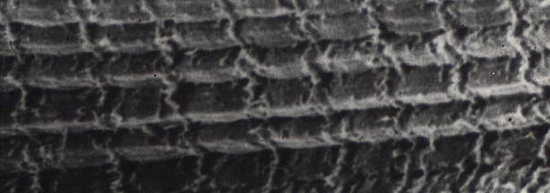
Transmission electron microscopy image of the sarcomere, with... | Download Scientific Diagram

Polarization-resolved microscopy reveals a muscle myosin motor-independent  mechanism of molecular actin ordering during sarcomere maturation | PLOS  Biology
Polarization-resolved microscopy reveals a muscle myosin motor-independent mechanism of molecular actin ordering during sarcomere maturation | PLOS Biology

Muscle structure – muscle under the microscope — Science Learning Hub